
Age-related macular degeneration can be diagnosed during an eye exam. Your ophthalmologist may have you look at an Amsler grid. This 20x 20 square grid that looks like graph paper helps you identify any blurry, distorted, or missing areas in your central field of vision.
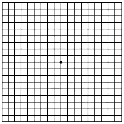
Normal Amsler Grid
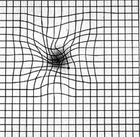
Amsler Grid in Macular Degeneration
with dark spot and distortion
On the dilated exam, your eye doctor will look for drusen, named after the German word for pebble. Drusen look like small yellowish-white stones underneath the retina. Drusen are the hallmark of macular degeneration and represent oxidative stress and inflammation in the eye, two of the root causes of macular degeneration. The presence of drusen on their own are classified as dry macular degeneration.
Drusen “pebble” deposits may increase in size and number as macular degeneration progresses. If the inflammation continues chronically, then new blood vessels grow under the retina and leak, in which case macular degeneration has transformed into the wet stage. I like to call this stage of macular degeneration “leaky eye” syndrome.
You may be familiar with the concepts known as leaky gut, leaky brain, or leaky heart. Similar to the other “leaky” syndromes, wet macular degeneration is a form of “leaky eye” syndrome. When macular degeneration becomes wet, blood, fluid, and proteins leak underneath and into the retina, leading to significant vision loss.
Drusen and fluid in or underneath the retina can be detected on a non-invasive imaging test called Optical Coherence Tomography (OCT).
Your ophthalmologist may also use fluorescein angiography to see what is going on with your retina. Fluorescein, a yellow dye is injected into a vein, typically in your arm. This dye then travels throughout your blood vessels and uses a camera to take images of the retina. This can detect if there are abnormal new blood vessels under the retina.
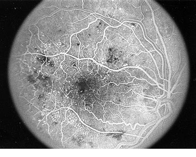
Another way to check the blood vessels in and under the retina is by using an optical coherence tomography angiography (OCTA). This process is similar to fluorescein angiography but without the use of a dye.
Click HERE to learn more about macular degeneration and ways you can prevent vision loss.
Adapted from AAO.org
Images Source: AAO.org
Whether the practice of Rudrani Banik, MD is the first ophthalmology office you are visiting for eye treatment, or simply the last one, Dr. Banik will make sure she does everything in her power to find an effective treatment to help you see better.